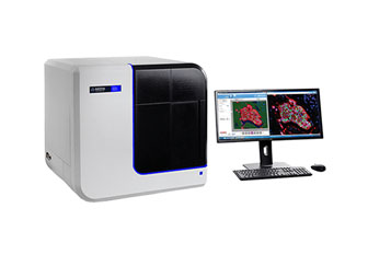
PhenoImager® HT 2.0
PhenoImager® HT 2.0
Fast-Track Spatial Signatures with High-Throughput
Imaging
PhenoImager HT
2.0 is the fastest and most widely adopted spatial imaging platform in
translational research today. Purpose-built for scalable biomarker discovery,
it empowers researchers to visualize, quantify, and decode spatial
phenotypes across hundreds of tissue samples each week—with unmatched speed
and clarity.
Spatial Signatures
The Next Generation of Predictive Biomarkers
Spatial
signatures combine cell type, phenotype, and location into a tissue-based
biomarker that drives precision insights. PhenoImager HT 2.0 is designed to
accelerate their development through whole-slide multispectral imaging, spectral
unmixing, and automated high-throughput workflows.
PhenoImager HT
2.0 – Spatial Signatures at Your Scale
- Brightfield and fluorescence imaging (up to 9 colours)
- 6-plex whole-slide scans in less than 12 minutes
- Onboard spectral unmixing performed in parallel with scanning
- 400+ slides/week throughput with walk-away
automation
- Seamless integration with PhenoCode™ Signature Panels
Technology Highlights
High-Throughput Imaging Powered by Multispectral
Precision
PhenoImager HT
2.0 combines imaging speed with exceptional clarity using Akoya’s patented MSI
technology. This allows precise phenotyping even in dense, auto fluorescent tissues.
- Detect up to 9 markers per slide with clear signal separation
- Perform full-slide, high-resolution imaging in minutes
- Reduce background and autofluorescence for clean data
- Ideal for FFPE, fresh frozen, and challenging tissues
Workflow Integration
Effortless Scalability for Every Lab
From discovery
labs to regulated clinical sites, PhenoImager HT 2.0 fits seamlessly into
existing research infrastructure.
- Compatible with PhenoCode™ Panels & Signature Panels
- Works with Phenochart™, HALO®, and AI-powered analytics tools
- Scalable to meet small study or high-volume cohort demands
- Supports batch analysis and retrospective studies using TMAs
Applications
Built for Precision Research Across Disease Areas
PhenoImager HT
2.0 supports a wide spectrum of biological and clinical research by delivering
high-content spatial information at scale. Its applications include:
Cancer &
Tumor Microenvironment
- Immune cell infiltration and checkpoint marker analysis
- Spatial mapping of tumor–immune interactions
- Immunotherapy response prediction
Immunology
& Inflammation
- Immune subset profiling in tissue context
- Chronic inflammation and autoimmunity studies
- Vaccine response evaluation
Neuroscience
- Microglia, astrocyte, and neuron spatial distribution
- Brain region mapping and neuroinflammation
- Neuropathology and neurodegeneration studies
Infectious
Disease
- Spatial distribution of pathogen and host markers
- Tissue-level immune response profiling
- Lung, gut, and mucosal barrier investigations
Fibrosis &
Tissue Remodeling
- ECM and stromal marker analysis
- Spatial dynamics in organ fibrosis
- Wound healing, regeneration, and chronic injury
Translational
& Clinical Research
- Retrospective biomarker validation in tissue microarrays (TMAs)
- Prognostic and predictive spatial signature development
- Large-cohort slide scanning with reproducibility
- Versatile: High Quality 40X Resolution Ideal for RNA-FISH or Multispectral Bright field imaging.